Patellofemoral Instability
Patellofemoral Instability results from one or more dislocations or partial dislocations, also called subluxations. This misalignment can damage the underlying soft structures such as muscles and ligaments that hold the knee in place. Once damaged, these soft structures are unable to keep the patella (knee cap) in position.
Signs and symptoms of Patellofemoral Instability can include the following:
- Pain, especially when standing up from a sitting position
- Feeling of unsteadiness or tendency of the knee to “give way” or “buckle”
- Recurrent Subluxation: When the kneecap slips partially out of place repeatedly
- Recurrent Dislocation: When the kneecap slips all the way out of position repeatedly
- Severe pain, swelling and bruising of the knee immediately following subluxation or dislocation
- Visible deformity and loss of function of the knee often occurs after subluxation or dislocation
- Sensation changes such as numbness or even partial paralysis can occur below the dislocation as a result of pressure on nerves and blood vessels
Causes
Patellofemoral Instability can be caused by a number of factors that affect the way the patella moves along the groove of the femur (trochlear groove) when the leg is bent or straightened. The patella normally moves up and down with a slight tilt without touching the other knee bones. In Patellofemoral Instability the patella does not maintain its normal path of movement and can slip out of the trochlear groove either partially (subluxation) or completely (dislocation).
Diagnosis
Evaluating the source of Patellofemoral Instability is critical in determining your treatment options for relief of the instability. Your physician will perform the following:
- Medical History
- Physical Examination
- Diagnostic studies such as X-rays, CT scan, or MRI
The goal of conservative treatment for Patellofemoral Instability is to restore full range of motion by restoring the normal tracking pathway of the patella during flexion and extension of the knee. Treatment options may include closed reduction, pain medications, rest, ice, physical therapy, orthotics, and bracing. Surgical treatment of Patellofemoral Instability is sometimes necessary to help return the patella to a normal tracking path when conservative treatment options are unsuccessful.
Patellofemoral Realignment
Medial patellofemoral ligament realignment is a surgical procedure indicated in patients with severe patellar instability. Medial patellofemoral ligament is a band of tissue that extends from the femoral medial epicondyle to the superior aspect of the patella. Medial patellofemoral ligament is the major ligament which stabilizes the patella and helps in preventing patellar subluxation (partial dislocation) or dislocation. This ligament can rupture or get damaged due to lateral dislocation of the patella. Dislocation can be caused by direct blow to the knee, twisting injury to the lower leg, strong muscle contraction, or because of a congenital abnormality such as shallow or malformed joint surfaces.
Medial patellofemoral ligament realignment using autogenous tissue grafts is done by following the basic principles:
- Graft Selection: Strong and stiff graft should be selected
- Location: The graft should be located isometrically
- Correct tension: The tension set in the graft should be appropriate
- Secure Fixation: Stable fixation of the graft should be achieved
- Avoid condylar rubbing or impingement: The graft should not rub against condyle or cause impingement
Surgical Technique
The surgical procedure of medial patellofemoral ligament realignment involves the following steps:
Graft Selection and Harvest: Your surgeon will make a 4-6 cm skin incision over your knee, at the midpoint between the medial epicondyle and the medial aspect of the patella (knee cap). The underlying subcutaneous fat and fascia are cut apart to expose the adductor tendon. The tendon is then stripped using a tendon stripper and its free end is sutured. The diameter of the tendon graft is measured using a sizer.
Alternatively, a graft can be harvested from the quadriceps tendon.
Location of the femoral isometric point: The graft should be placed isometrically to prevent it from overstretching and causing a failure, during joint movements. A transverse hole, measuring 2.5 mm, is made through the patella. Then a small incision is made over the lateral side of the patella and a strand of Vicryl suture material is inserted through the hole. Over this strand, a 2.5 mm Kirschner wire (K-wire) is passed and then inserted into the bone beside the medial epicondyle.
An instrument called pneumatic isometer is inserted into the hole made in the patella and the Vicryl isometric measurement suture material is also passed along. The knee is taken through its full range of motion and any changes happening in the length between the medial epicondylar K-wire and the medial aspect of the patella is recorded on the isometer. The position of the K-wire will be adjusted until no deviations are read on the isometer during full range of motion. Once the isometric point is identified, a tunnel is drilled starting from the insertion of the adductor tendon uptil the isometric point is reached. The graft is pulled through this tunnel, then exits at the medial condyle and again passed through another tunnel that is made through the patella.
Correct tension: The tension is set in the graft with your knee flexed upto 90º and the tension should be appropriate enough to control lateral excursion.
Secure fixation: After bringing the tendon graft from the medial to the lateral side through the bone tunnel, it turned onto the front surface of the patella where it is sutured.
Avoid condylar rubbing and impingement: After graft fixation, the range of motion is checked to make sure there are no restrictions in patellar or knee movements. The graft should not impinge or rub against the medial femoral condyle. If it is detected, the graft is replaced into proper position.
Post-operative care
A knee brace should be used during walking in the first 3-6 weeks after surgery. Avoid climbing stairs, squatting and stretching your leg until there is adequate healing of the tendon. Rehabilitation exercises, continuous passive motion and active exercises will be recommended.
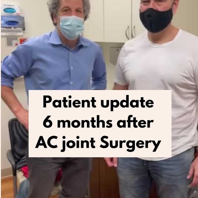
Contact our office today for a consultation with Dr. Steven Struhl of Shoulders & Knees.